|
 Use headphones for best audio quality. Use headphones for best audio quality.
|
“You only see what you look for.” In the physical examination of the heart, the adage becomes “You only hear what you listen for.” A very experienced clinician may listen to a polyglot of noises, compare them to an internal matrix, and make a correct diagnosis with, “that sounds like….” For those of us who are mere mortals, proper auscultation requires that we listen carefully and thoughtfully to one thing at a time. We must seek out normal and abnormal sounds and murmurs rather than waiting for them to announce themselves. Perhaps the most difficult skill to acquire is separating distinct sounds and describing their timing and character. A loud systolic murmur may be easy to identify. By habit, we almost automatically place all action in systole, but to describe when in systole the murmur occurs requires a bit of practice. You must listen to the murmur and only the murmur in several locations. You then listen to the distinct and separate normal or expected sounds to determine the timing as midsystolic or holosystolic. Perhaps it is continuous and not really just a systolic murmur. You must seek out additional sounds that may be hiding within the murmur, for example, an ejection sound of bicuspid aortic valve. Without this effort to listen for distinct sounds and their timing, a correct diagnosis from physical examination will be elusive.
When you examine a sound or murmur, ask 4 questions to help you determine their origin. Ask when, where, with whom, and what pitch. 1) When do you hear the sound? During systole, diastole, early, mid, or late? 2) Where can you hear the sound or murmur most easily? 3) With whom is perhaps the most important question. Where else (at what other locations) can you hear the sound or murmur (radiation)? What other sounds or murmurs are also present? Many times a diagnosis rests not on the character of a particular sound but on the company it keeps. For example, you may notice a similarity between the widely radiating pericardial knock and an opening snap of mild mitral stenosis. When you encounter the extra sound, careful examination in the left lateral decubitus position will expose the murmur of mitral stenosis. Meanwhile, close inspection of the precordium and neck veins may uncover the company kept by a pericardial knock. 4) The fourth question is about pitch; murmurs or sounds in the high frequency range such as valve closure sounds, clicks, or the murmur of mitral regurgitation are heard best with the diaphragm. This does not mean that they cannot be heard with the bell, but they are louder or more easily appreciated with the diaphragm. Conversely, low frequency sounds or murmurs such as the S3 or murmur of mitral stenosis are more easily heard with the bell.
Diastolic sounds and murmurs represent a greater listening challenge than events in systole. As stated earlier, our imagination moves all action to systole. Therefore, our attention is centered there. We may easily overlook diastolic sounds or murmurs or mistakenly think they occur in systole. You can usually determine timing at the bedside once you have identified the first and second heart sounds. Alternatively, simultaneous palpation of the carotid pulse may be helpful. Four diastolic sounds are presented here on this web preview as an example. They are the third heart sound–the opening snap of rheumatic mitral stenosis, the S3 of left ventricular failure, the pericardial knock, and the tumor plop of left atrial myxoma. Although the sounds bear some similarity, if you ask the 4 questions and use the simultaneous ECG tracing, you should be able to distinguish each one.
Mitral stenosis is associated with four auscultatory findings: loud S1, loud P2, an opening snap, and a diastolic murmur. Only the murmur is consistent. The loudness of S1 and S2 are affected by the patient’s body habitus, valve mobility, and pulmonary artery pressure. The still mobile mitral valve that has been damaged by rheumatic fever creates a high frequency sound in early diastole. As left ventricular pressure falls below left atrial pressure, the mitral valve is thrust open. When reaching its maximal excursion due to the limits placed by tethering and fibrosis, the valve comes to a sudden halt. Its thin and relatively inelastic structure creates a high frequency vibration that you can hear best with the diaphragm. The opening snap requires a relatively mobile and non-calcified valve. In many patients with severe and long-standing rheumatic mitral stenosis, calcification of the valve reduces mobility and the OS disappears.
You can hear the opening snap near the cardiac apex, but more easily appreciate it along the lower left sternal border. Its proximity to aortic valve closure may create confusion with a widely split second sound. You may find the sound less confusing if you inch upward along the left sternum border and note a decline in the intensity of the early diastolic sound in conjunction with audible splitting of the second sound with inspiration. It is important to listen carefully to a mitral opening snap so you can identify a still mobile mitral valve and estimate the severity of mitral stenosis. As the severity of mitral stenosis increases, left atrial pressure rises. Rising left atrial pressure results in mitral valve opening at earlier time points along the decline of left ventricular pressure during relaxation. Therefore, the time from aortic valve closure sound to opening snap (often referred to as the A2-OS) offers us a method for estimating the severity of the disease. Consider also that A2-OS may be influenced by heart rate, impaired left ventricular relaxation, and concurrent mitral regurgitation.
 |
|
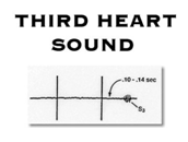
The S3 is a low frequency sound, generally heard from 120-160 milliseconds after the aortic valve closure sound. In order to find an S3, place the patient in the left lateral decubitus position and identify the apical impulse. Place the bell lightly against the skin surface over the palpated apical impulse. In childhood and physically active young adults, the third heart sound is felt to arise from vigorous elastic recoil of the left ventricle in early diastole, causing the heart to impact the chest wall. Here, the S3 is a normal finding. In adults, the third heart sound is a physical sign specific for left ventricular failure. It is heard predominantly in dilated hypocontractile left ventricles, but may also be heard in the setting of severe mitral regurgitation. In left ventricular failure, the origin of the third heart sound is thought to be the sudden cessation of early, rapid left ventricular filling impelled by high left atrial pressure. This low frequency sound approaches the limits of human hearing and may not radiate widely from the apical impulse. When you are uncertain about the presence of an S3, a brief period of exercise for the patient will augment the amplitude, making detection easier.
 |
|
|
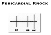
The early diastolic sound of pericardial constriction occurs slightly earlier than the average third heart sound. Its frequency is somewhat higher, allowing you to hear it throughout the precordium using the diaphragm and the bell. Commom causes of pericardial constriction include prior cardiac surgery and uremic pericarditis. Outside the United States, the most common cause is tuberculous pericarditis. Idiopathic pericarditis and prior bacterial pericarditis are uncommon causes. Associated physical findings include elevated jugular venous pressure with a rapid Y-descent. The rapid descent of the jugular veins can usually be seen to occur in conjunction with or just following the pericardial knock. Patients are frequently in atrial fibrillation and have physical findings of right-sided heart failure. Often, edema is less prominent than ascites and there may be associated proximal muscle wasting as these patients are frequently symptomatic for several years before the diagnosis is established. Kussmaul’s sign, an inspiratory rise in jugular venous pressure, may be seen in approximately 30-40% of patients with constrictive pericarditis.
 |
|
|
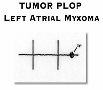
A large left atrial myxoma produces a diastolic sound that is referred to as a tumor plop. This sound arises from obstruction to ventricular in-flow that occurs as the tumor comes to rest over the mitral annulus. It is the functional equivalent of mitral valve stenosis and may be associated with a low frequency diastolic murmur. Over time, damage to the mitral valve may occur resulting in a concurrent regurgitant murmur. Although similar in timing to mitral valve stenosis, the tumor plop differs from the mitral opening snap in that it is a low frequency sound and is thus heard best with the bell. Diagnosing atrial myxoma is similar to diagnosing the pericardial knock. It is as much about the company it keeps as it is about the characteristics of the sounds you hear. Atrial myxoma is slightly more common in women and there is generally no prior history of rheumatic fever. Patients may present with dysnea, orthopnea, palpitation, fever, and embolic phenomenon. An abnormal ANA may be seen in conjunction with atrial myxoma.
Return to
Heart Sounds Auscultation