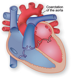
The aorta is the artery that carries oxygen-rich blood from the lower-left chamber (the left ventricle) to the rest of the body. Coarctation of the aorta (also called CoA) means that one area of the aorta is abnormally narrow.
 The arteries that carry blood to different parts of the body branch off of the aorta. In children with coarctation, the arteries that branch off before the narrow point receive more blood than the arteries that branch off after it. This increases blood pressure in the arteries that supply blood to the head and arms and decreases blood flow to the legs. The heart is forced to work harder, causing it to get larger. In time, this extra work leads to heart failure.
What are the symptoms?
- Blood pressure that is higher in the arms than in the legs
- Weak pulse or no pulse in the groin area
- Cold legs or feet
- Nosebleeds
- Dizziness
- Fainting
- Leg cramps with exercise
Symptoms usually do not appear until about a week after birth. In a newborn, a narrowed aorta may not be noticed until the ductus arteriosus closes in the days after birth. The narrowing sometimes appears along with other conditions like septal defects and valve problems.
Sometimes coarctation is not diagnosed until late childhood or the teenage years.
With coarctation of the aorta, early detection is important. Coarctation can cause high blood pressure. The longer it goes untreated, the more likely it is that the child will have high blood pressure throughout his or her life.
How is it treated?
Surgery is recommended in most cases. Surgery usually involves removing the narrowed part of the aorta. If this part is small, the two free ends of the aorta can be reconnected after the narrow part is removed. Otherwise, a tube of fabric, a fabric patch, or a flap of tissue may be used to connect the two ends.
To widen the narrowed segment without surgery, doctors may use a catheterization procedure called balloon angioplasty.
Also, doctors use wire-mesh devices called stents to support the aorta and keep it open. These stents are implanted during an angioplasty procedure.
Return to main topic: Congenital Heart Disease
See on other sites:
MedlinePlus
https://medlineplus.gov/ency/article/000191.htm
Coarctation of the Aorta
American Heart Association
www.heart.org/HEARTORG/Conditions/CongenitalHeartDefects/
AboutCongenitalHeartDefects/Coarctation-of-the-Aorta-CoA_UCM_307022_Article.jsp
Coarctation of the Aorta (CoA)
Texas Adult Congenital Heart Center (TACH)
www.bcm.edu/healthcare/care-centers/congenital-heart
This Baylor College of Medicine program enables patients with congenital heart disease to receive a seamless continuation of care from birth to old age.
Updated August 2016


