Intravascular ultrasound (IVUS) or intravascular echocardiography is a combination of echocardiography and a procedure called cardiac catheterization. IVUS uses sound waves to produce an image of the coronary arteries and to see their condition. The sound waves travel through a tube called a catheter. The catheter is threaded through an artery and into your heart. This test lets doctors look inside your blood vessels.
IVUS is rarely done alone or as a strictly diagnostic procedure. It is usually done at the same time that a percutaneous coronary intervention, such as angioplasty, is being performed.
How does it work?
|

|
|
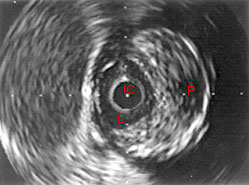
IVUS image of inside a coronary artery.
IC=IVUS catheter, L=lumen, P=plaque
|
|
IVUS uses high-frequency sound waves (also called ultrasound) that can provide a moving picture of your heart. The pictures come from inside the heart rather than through the chest wall. The sound waves are sent with a device called a transducer. The transducer is attached to the end of a catheter, which is threaded through an artery and into your heart. The sound waves bounce off of the walls of the artery and return to the transducer as echoes. The echoes are converted into images on a television monitor to produce a picture of your coronary arteries and other vessels in your body.
See also on this site:
See on other sites:
MedlinePlus
https://medlineplus.gov/ency/article/007266.htm Intravascular ultrasound
Updated August 2016


