A nuclear ventriculography scan is a test using radioisotope dye that tracks blood flow through your heart during rest, exercise, or both. The test can tell doctors how well the heart is pumping blood and if it is working harder to make up for one or more blocked arteries. This scan is also very useful for finding your "ejection fraction," which is the percentage of blood that is pumped out of your heart's lower chambers (called the ventricles) with each heartbeat. This test is also called radionuclide ventriculography (RVG, RNV) or multiple-gated acquisition (MUGA) scan.
How does it work?
|
|
|
Diagnostic image from MUGA scan.
|
A MUGA scan makes use of a radioactive substance that is injected into your bloodstream. The radioactive substance "tags" or "labels" the red blood cells in your blood. This substance is safe and will not harm your blood or organs. Doctors will then use a gamma-ray camera to take pictures of your heart as the "tagged" red blood cells circulate.
What should I expect?
No special preparation is needed before you have a resting scan. If your doctor has ordered an exercise scan, do not eat or drink anything (you may only have water) after midnight the night before the test.
If you think you may be pregnant, talk to your doctor before you schedule the test.
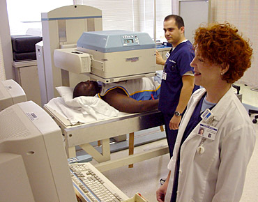
A technician will clean certain areas on your chest so that he or she can place small metal disks called electrodes on those areas. The electrodes have wires called leads, which are attached to a nuclear imaging computer. Then the technician will give you 2 injections: the first injection prepares the red blood cells, and the second is used to "label" the red blood cells. The technician will ask you to lie down on a small examination table, which has a special camera around it. Next, the technician will take a number of pictures of your heart with the gamma-ray camera. If your doctor only ordered a resting scan, this would be the end of the test.
 |
|
Technicians conduct MUGA scan.
|
If your doctor ordered an exercise scan, you will be moved to a different examination table. When you lie down, there will be pedals at the end of the bed. You will put your feet in the pedals and, while still lying down, begin to pedal as if you were riding a bicycle. Using the gamma-ray camera, the technician will take a number of pictures of your heart. Your doctor may also be present to look at the pictures of your heart during the test.
After the test, you may feel tired, but you will be allowed to resume your normal activities as soon as you are done with the test. The harmless radioactive substance will leave the body within 2 or 3 days. Women who are pregnant or breastfeeding should not have a MUGA scan.
See on other sites:
American Heart Association
www.heart.org/HEARTORG/Conditions/HeartAttack/SymptomsDiagnosisofHeartAttack/Radionuclide-Ventriculography-or-Radionuclide-Angiography-MUGA-Scan_UCM_446354_Article.jsp
Radionuclide Ventriculography (MUGA Scan)
Updated August 2016


