Imaging Tests
Computed tomography (CT or CAT scan) is an X-ray technique that uses a computer to create cross-sectional (or slice-like) pictures of your brain. A CT scan can show whether you have had a stroke. A CT scan can also help doctors identify the type of stroke you have had—ischemic (the result of a blockage) or hemorrhagic (the result of bleeding).
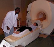
Magnetic resonance imaging (MRI) is an imaging procedure that uses a magnetic field to cause a change in the behavior of your brain cells. Your cells react to the field of energy in the form of radio signals, which a computer reads and turns into an extremely accurate image of your brain. An MRI can reveal the presence, location, and size of an aneurysm or malformation in the arteries and veins, which can cause a hemorrhagic stroke.
Ultrafast MRI Gives Rapid Diagnosis
A traditional MRI takes about 30 minutes to complete. A new, ultrafast MRI uses faster shifting and stronger magnetic fields to give a picture of the brain in 15 minutes or less. This faster way of diagnosing stroke may make a difference in the care of stroke patients. Ultrafast MRI lets doctors avoid angiography (a test to see inside the blood vessels) in patients who do not need it. This is important because patients can get clot-dissolving medicines sooner, possibly preventing long-term complications such as muscle weakness and paralysis. Doctors must give clot-dissolving medicines within 3 hours of the start of stroke symptoms.
Angiography is an X-ray technique in which dye is used to study your blood circulation. The procedure can show blocked blood vessels and areas of the brain that have been without blood.
Electrical Activity Measurements
An electroencephalogram (EEG) gives doctors a record of the electrical impulses produced by your brain. Small metal disks (called electrodes) are placed on your scalp. The electrodes pick up your brain's electrical activity in the form of impulses, which are put on paper. The intensity, duration, frequency, and location of the impulses can tell your doctor a lot about your brain function.
An evoked response test measures your brain's ability to process and react to your environment. For example, flashing a light or checkerboard pattern in front of your eyes will evoke a visual response. Making a sound in your ear will get an auditory response. Or, electrically stimulating one of the nerves in your arm or leg will provide a bodily response. The responses can tell doctors if there are abnormal areas in your brain.
Blood Flow Tests
Doppler ultrasound uses sound waves to examine blood flow in your carotid arteries. The carotid arteries are the arteries in your neck that carry blood to your brain. The sound waves used for Doppler ultrasound are delivered through a device called a transducer. When the transducer is placed over the carotid artery, the sound waves travel through your neck and bounce off the moving blood cells as an echo. These echoes are converted to an image on a television monitor. The changes in frequency are related to the speed of blood cells, which, in turn, is related to blood flow. These changes may indicate a narrowing or blockage of the carotid artery.
Ultrasound imaging may also be used to determine the thickness of the walls of the carotid artery. This may help doctors predict heart attacks and strokes in older adults, according to a report in the New England Journal of Medicine. Researchers with the National Heart, Lung, and Blood Institute found that older adults are at increased risk for heart attack or stroke if ultrasound shows a thickening of the carotid arteries. In the future, use of ultrasound may let doctors provide aggressive treatment earlier.
In carotid phonoangiography, a sensitive microphone is placed on the neck to record the sound of the blood flow in the carotid artery. In a normal artery, blood flows smoothly. But if there is a blockage, the blood flow will sound rough and turbulent. This turbulent blood flow is called a bruit (pronounced bru-ee). A bruit tells doctors that there is a blockage in the carotid artery.
Return to main topic: Stroke
Updated August 2016