The heart weighs between 7 and 15 ounces (200 to 425 grams) and is a little larger than the size of your fist. By the end of a long life, a person's heart may have beat (expanded and contracted) more than 3.5 billion times. In fact, each day, the average heart beats 100,000 times, pumping about 2,000 gallons (7,571 liters) of blood.

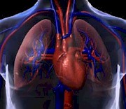
Your heart is located between your lungs in the middle of your chest, behind and slightly to the left of your breastbone (sternum). A double-layered membrane called the pericardium surrounds your heart like a sac. The outer layer of the pericardium surrounds the roots of your heart's major blood vessels and is attached by ligaments to your spinal column, diaphragm, and other parts of your body. The inner layer of the pericardium is attached to the heart muscle. A coating of fluid separates the two layers of membrane, letting the heart move as it beats.
Your heart has 4 chambers. The upper chambers are called the left and right atria, and the lower chambers are called the left and right ventricles. A wall of muscle called the septum separates the left and right atria and the left and right ventricles. The left ventricle is the largest and strongest chamber in your heart. The left ventricle's chamber walls are only about a half-inch thick, but they have enough force to push blood through the aortic valve and into your body.
The Heart Valves (illustration)
Four valves regulate blood flow through your heart:
- The tricuspid valve regulates blood flow between the right atrium and right ventricle.
- The pulmonary valve controls blood flow from the right ventricle into the pulmonary arteries, which carry blood to your lungs to pick up oxygen.
- The mitral valve lets oxygen-rich blood from your lungs pass from the left atrium into the left ventricle.
- The aortic valve opens the way for oxygen-rich blood to pass from the left ventricle into the aorta, your body's largest artery.
See also on this site: The Heartbeat
The Conduction System (illustration)
Electrical impulses from your heart muscle (the myocardium) cause your heart to contract. This electrical signal begins in the sinoatrial (SA) node, located at the top of the right atrium. The SA node is sometimes called the heart's "natural pacemaker." An electrical impulse from this natural pacemaker travels through the muscle fibers of the atria and ventricles, causing them to contract. Although the SA node sends electrical impulses at a certain rate, your heart rate may still change depending on physical demands, stress, or hormonal factors.
The Circulatory System (illustration)
The heart and circulatory system make up your cardiovascular system. Your heart works as a pump that pushes blood to the organs, tissues, and cells of your body. Blood delivers oxygen and nutrients to every cell and removes the carbon dioxide and waste products made by those cells. Blood is carried from your heart to the rest of your body through a complex network of arteries, arterioles, and capillaries. Blood is returned to your heart through venules and veins. If all the vessels of this network in your body were laid end-to-end, they would extend for about 60,000 miles (more than 96,500 kilometers), which is far enough to circle the earth more than twice!
See also on other sites:
MedlinePlus
https://medlineplus.gov/ency/imagepages/8672.htm
Normal heart anatomy (cut section illustration)
Updated August 2016